Deep Vein Thrombosis (DVT)
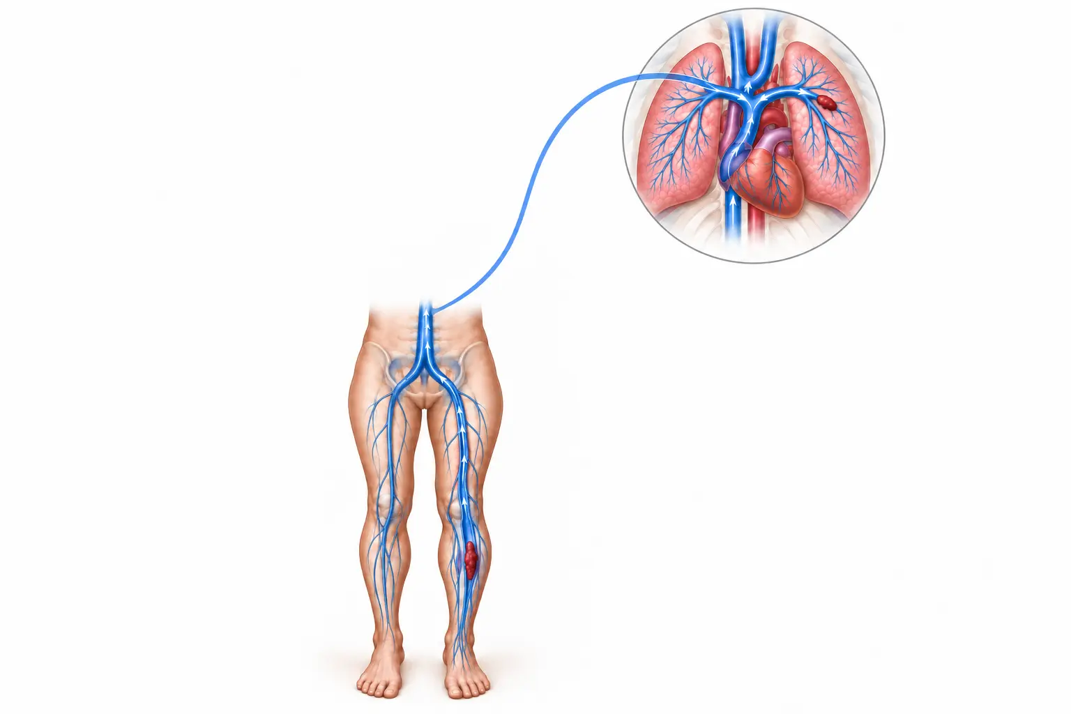
Deep vein thrombosis (DVT) is a blood clot in a deep vein, most often in the leg. It may cause sudden swelling, pain, warmth or colour change on one side, but some people have few symptoms. A suspected DVT needs prompt medical assessment because part of the clot can travel to the lungs and cause a pulmonary embolism.
Routine appointment contact is not an emergency service.
What is DVT?
A DVT forms when blood clots within a deep vein. Lower-limb DVT may involve veins below the knee, above the knee, or the pelvic veins. The clot can obstruct venous return and damage vein valves. It can also break away and travel to the lungs, producing a pulmonary embolism. DVT and pulmonary embolism together are called venous thromboembolism (VTE).
DVT is different from a clot in a superficial varicose vein, although either condition requires proper assessment when symptoms are new or extensive.
Symptoms that may suggest DVT
- New swelling affecting one leg more than the other
- Calf or thigh pain, tenderness, tightness or heaviness
- Warmth, redness or darker discolouration of the affected limb
- Swelling of the whole leg in a more extensive thrombosis
- New prominent superficial veins
- Unexplained limb symptoms after surgery, hospitalisation, prolonged immobility or long travel
Symptoms alone cannot confirm or exclude DVT. Muscle injury, cellulitis, a ruptured Baker’s cyst, lymphatic swelling, venous insufficiency and other conditions can look similar. Some DVTs produce little obvious swelling or pain.
Who is at increased risk?
Risk is increased by:
- Recent major surgery, trauma, fracture or hospital admission
- Reduced mobility, limb immobilisation or prolonged bed rest
- Active cancer and some cancer treatments
- Pregnancy and the first weeks after delivery
- Oestrogen-containing contraception or hormone therapy
- A previous DVT or pulmonary embolism
- Increasing age, obesity and some chronic medical illnesses
- An inherited or acquired tendency to clot in selected patients
- Long journeys, particularly when combined with other risk factors
A DVT may also occur without an obvious provoking factor. Risk factors guide assessment but do not establish the diagnosis.
How suspected DVT is assessed
Clinical assessment
The clinician reviews the onset and distribution of symptoms, recent risk factors, medication and bleeding risk, and examines both legs. A validated clinical-probability pathway may be used to decide which tests are needed and how urgently.
D-dimer blood test
D-dimer can help exclude DVT in appropriately selected people with a low or unlikely clinical probability. A positive result does not prove DVT because many other conditions can increase D-dimer. It is not a substitute for imaging when clinical probability is high.
Venous duplex ultrasound
Compression ultrasound with Doppler is the main imaging test for suspected lower-limb DVT. It assesses whether the deep veins compress normally and whether a clot is present. If the first scan is negative but clinical concern remains, repeat or additional imaging may be required according to the diagnostic pathway.
Additional imaging
CT venography, MR venography or other imaging may occasionally be needed when pelvic or iliac-vein thrombosis is suspected, ultrasound is inconclusive, or an intervention is being planned.
Treatment is individualised
Treatment depends on the clot’s location and extent, symptoms, provoking factors, kidney and liver function, pregnancy or cancer status, interacting medicines and bleeding risk.
Anticoagulation
Anticoagulants are the main treatment for most confirmed DVTs. They reduce further clot formation while the body gradually remodels the existing clot. The medicine, dose and monitoring plan must be prescribed for the individual patient.
For confirmed proximal DVT, treatment commonly includes at least three months of anticoagulation. Whether treatment stops or continues beyond the initial course depends on whether the event was provoked by a temporary factor, whether risk factors persist, whether the DVT was unprovoked or recurrent, and the balance between recurrence and bleeding risk.
Selected isolated distal calf DVTs may be managed with anticoagulation or structured ultrasound surveillance depending on symptoms and risk of extension. This decision requires clinician assessment.
Do not start, stop or change an anticoagulant based on website information. Seek urgent advice for major bleeding, black stools, vomiting blood, coughing blood, severe persistent headache, significant head injury or other concerning symptoms while taking anticoagulation.
Walking and daily activity
Once treatment has started and the patient is clinically stable, normal walking is generally encouraged rather than routine bed rest. Activity should be adapted to pain, swelling, cardiopulmonary symptoms and any separate medical or surgical restriction.
Compression
Compression stockings are not routinely prescribed solely to prevent post-thrombotic syndrome after every DVT. They may be considered for selected patients to control persistent swelling or discomfort after arterial circulation, fit and contraindications have been assessed.
Catheter-directed treatment or thrombectomy
Most DVTs do not require clot-removal procedures. Catheter-directed thrombolysis, pharmacomechanical treatment or venous thrombectomy may be considered in carefully selected patients with extensive, recent iliofemoral DVT, severe symptoms, good functional status and a low bleeding risk, or in limb-threatening venous obstruction. Potential benefit must be weighed against bleeding, access-site and procedure-related risks.
Inferior vena cava filter
An inferior vena cava (IVC) filter is not routine treatment when anticoagulation can be given. It may be considered in limited circumstances, such as an acute VTE with a contraindication to anticoagulation, with a plan for review and retrieval when appropriate.
Follow-up after DVT
Follow-up should confirm that anticoagulation is being taken safely, review bleeding or recurrence symptoms, address the provoking factor and decide the appropriate duration of treatment. Kidney function, weight, other medicines, planned procedures, pregnancy considerations and cancer status may affect management.
Routine thrombophilia testing is not required for every patient. Testing is considered selectively when the result could change management and should be timed so that the acute clot and anticoagulant treatment do not make interpretation misleading.
Post-thrombotic syndrome
Some people develop persistent swelling, heaviness, aching, skin pigmentation, eczema or, less commonly, venous ulceration after DVT. This is called post-thrombotic syndrome. Ongoing symptoms warrant review to exclude recurrent thrombosis and to plan symptom control, compression when suitable, exercise, weight management, skin care and further venous assessment when indicated.
When to arrange a vascular consultation
Arrange prompt assessment for new one-sided swelling, unexplained calf or thigh pain, or new symptoms after surgery, immobility, cancer treatment, pregnancy/postpartum or a previous VTE. A routine clinic appointment is also appropriate for persistent symptoms after a treated DVT, anticoagulation planning around procedures, or review of extensive iliofemoral thrombosis.
If pulmonary-embolism symptoms or severe whole-leg swelling with blue/dusky discolouration are present, go directly to an emergency department.
Consultation in Bengaluru
Consult Dr Siddharth Matad, Vascular & Endovascular Surgeon, at Siddharth Vascular Clinic, United Hospital, Jayanagar, Bengaluru.
For routine appointments, call or WhatsApp +91 97465 53250, or use the Appointments page.
For emergency symptoms, go to the nearest emergency department. Do not wait for a routine appointment or a phone/WhatsApp response.
Frequently asked questions
Can DVT occur without obvious swelling?
Yes. Some DVTs cause mild or atypical symptoms, and a clot cannot be ruled out only because swelling is absent. Clinical probability and appropriate testing determine whether imaging is needed.
Is every swollen leg caused by DVT?
No. Cellulitis, venous insufficiency, lymphoedema, injury, a ruptured Baker’s cyst and other conditions can cause similar symptoms. New one-sided swelling still requires timely assessment because examination alone may not reliably distinguish them.
How is DVT confirmed?
Venous duplex ultrasound is the main test. Clinical probability and, in selected patients, a D-dimer blood test help determine the diagnostic pathway. Repeat or additional imaging may be needed when concern remains despite an initial negative scan.
How long is anticoagulation required?
Confirmed proximal DVT is commonly treated for at least three months. Longer treatment may be advised when the event was unprovoked, recurrent or associated with a continuing risk factor. Bleeding risk and patient preferences are part of the decision.
Is walking safe after DVT treatment starts?
For most clinically stable patients receiving appropriate treatment, walking is encouraged as tolerated rather than routine bed rest. Individual restrictions may apply after surgery, with severe symptoms or if pulmonary embolism is suspected.
Does every DVT need clot removal or a stent?
No. Anticoagulation is the main treatment for most DVTs. Clot-removal procedures and venous stenting are considered only in selected patients after assessment of clot extent, symptoms, anatomy, timing and bleeding risk.
Can DVT come back?
Yes. Recurrence risk varies with the cause of the first event, persistent risk factors and whether the event was unprovoked. Follow-up helps determine treatment duration and future prevention.
When is DVT an emergency?
Sudden breathlessness, chest pain, coughing blood, fainting or collapse may indicate pulmonary embolism and requires immediate emergency care. Severe whole-leg swelling with marked pain and blue or dusky discolouration is also an emergency.
Vascular consultation in Bengaluru
Routine outpatient assessment is available with Dr Siddharth Matad, Vascular & Endovascular Surgeon, at Siddharth Vascular Clinic, United Hospital, Jayanagar, Bengaluru.
For emergency symptoms, go directly to the nearest hospital emergency department.