Non-Healing Leg and Foot Wounds
A non-healing leg or foot wound is one that persists, deteriorates or fails to make expected progress despite appropriate care. Healing depends on adequate blood supply, pressure relief and treatment of contributing problems such as venous hypertension, neuropathy, infection when present, oedema and poor glucose control—not dressings alone. Early assessment helps identify the dominant cause or combination of causes so that treatment can be directed appropriately.
Routine appointment contact is not an emergency service.
What constitutes a non-healing wound
There is no single time threshold that defines every non-healing wound. Concern increases when a wound persists, enlarges, deepens or fails to show expected progress despite appropriate care. Duration, location, depth, surrounding tissue, perfusion, pressure, infection and patient factors all matter. Many leg and foot wounds are multifactorial. In a diabetes-related foot ulcer, failure to reduce wound area substantially after four weeks of appropriate care is a reason to reassess vascular status and the overall treatment plan; it is not a universal definition for all wounds.
Why dressings alone may not solve the problem
Dressings manage the wound surface (moisture, protection, exudate). They cannot restore inadequate arterial perfusion, correct severe venous hypertension, relieve repetitive pressure on an insensate foot, eradicate deep infection, or reverse neuropathy. Without addressing the underlying drivers, even the best dressings often fail to achieve durable healing.
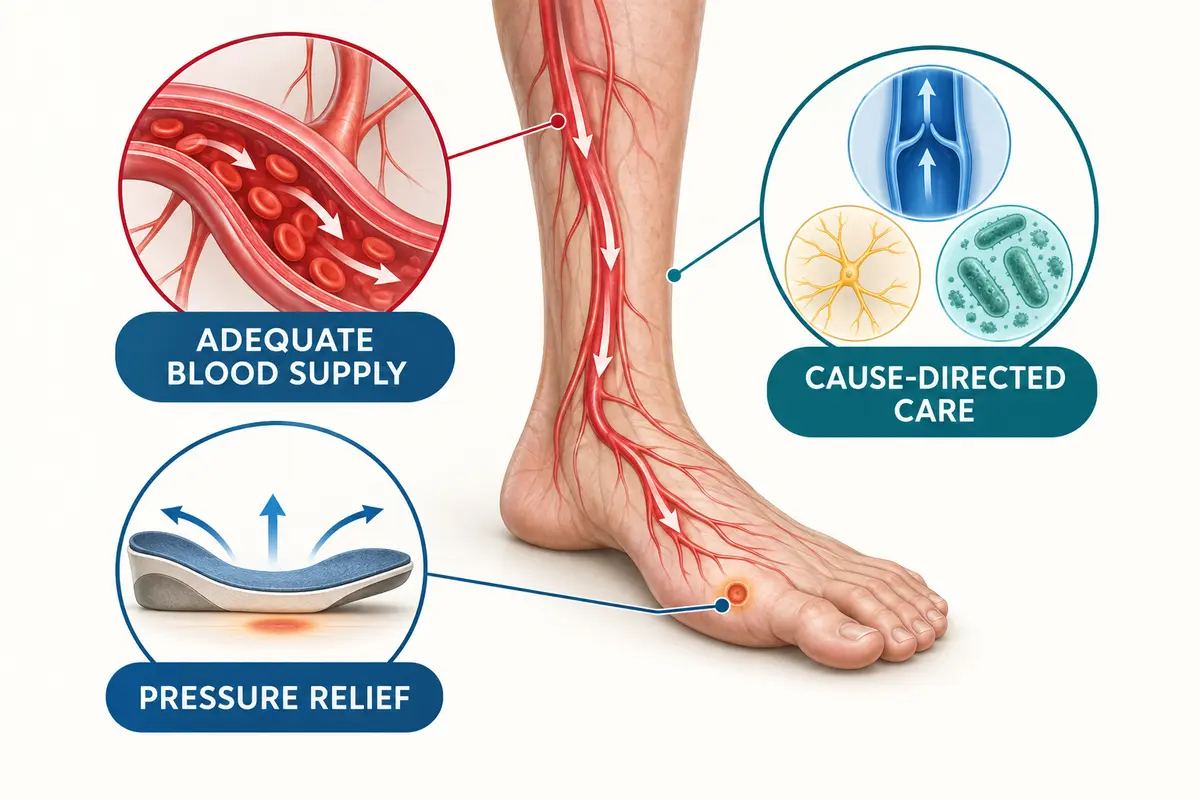
Major contributing causes
- Arterial insufficiency – reduced oxygenated blood flow from peripheral artery disease limits tissue repair and increases infection risk.
- Venous hypertension – sustained high pressure in the veins leads to oedema, skin changes and ulceration, commonly around the gaiter area.
- Diabetes and neuropathy – loss of protective sensation allows unnoticed trauma; metabolic factors and coexisting arterial disease may further impair healing. Diabetes-related microangiopathy should not be assumed to be the primary cause before other causes are assessed.
- Pressure – continuous or repetitive loading (especially plantar surfaces in neuropathy) causes tissue breakdown.
- Infection – local or deep soft-tissue infection and osteomyelitis can delay healing; not every non-healing wound is infected.
- Trauma – minor injury that fails to heal because of compromised circulation or sensation.
- Mixed disease – combination of arterial, venous and neuropathic factors is common and requires careful sequencing of care.
Symptoms and red flags
Common features include persistent open skin, surrounding redness or discolouration, swelling, drainage, odour, pain (or painless wounds in neuropathy), and callus. Red flags for urgent hospital assessment are listed in the emergency box above. Distinguish rapidly progressive infection (systemic illness + spreading erythema) from acute limb ischaemia (sudden cold, pale, pulseless, painful or numb limb).
Clinical assessment
Assessment begins with a full history (duration, trauma, diabetes control, previous vascular disease, smoking, footwear) and examination of both limbs, the wound bed, surrounding skin, oedema and footwear. Systemic features and cardiovascular risk factors are recorded.
Vascular examination and pulses
Palpation of femoral, popliteal, dorsalis pedis and posterior tibial pulses is performed. Absent or diminished pulses raise concern for significant arterial disease and prompt further physiological testing.
Neuropathy and diabetic-foot assessment
Protective sensation is tested (monofilament or equivalent). Foot deformity, callus, previous ulceration and footwear are evaluated. Patients with diabetes and neuropathy require structured foot examination even when the wound appears minor.
ABI and its limitations in diabetes/calcified arteries
Ankle-brachial index (ABI) compares ankle and arm systolic pressures. A low ABI supports the presence of PAD, but no single bedside value excludes PAD in a person with diabetes and a foot wound. Medial arterial calcification can make ankle arteries incompressible and produce a falsely elevated or unreliable ABI. Pedal Doppler waveforms, toe-brachial index and toe pressure are therefore interpreted together when appropriate.
Toe pressure and other physiological testing
Toe-brachial index or absolute toe pressure can add useful information about distal perfusion, particularly when ABI is unreliable. Transcutaneous oxygen pressure or skin perfusion pressure may be used selectively to estimate healing likelihood. These measurements inform probability; none guarantees healing or amputation.
Arterial and venous duplex ultrasound
Duplex ultrasound maps arterial stenoses/occlusions and assesses venous reflux or obstruction. It is non-invasive and guides decisions about further imaging or intervention.
When angiographic imaging may be needed
Catheter angiography, CT angiography or MR angiography is considered when revascularisation is planned or when non-invasive tests leave uncertainty about the extent and location of arterial disease. Imaging is not required for every non-healing wound.
Treatment principles
- Cause-directed wound care – identify and treat the dominant drivers (perfusion, venous pressure, pressure, infection, glycaemia).
- Debridement when appropriate – removal of non-viable tissue to prepare a healthy wound bed, taking ischaemia and anticoagulation into account.
- Infection assessment and treatment – clinical diagnosis of infection; microbiological sampling when indicated; systemic antibiotics only when infection is present.
- Pressure relief / offloading – redistribution or elimination of mechanical stress, especially for plantar diabetic-foot ulcers.
- Moisture balance – appropriate dressings chosen for exudate control and patient comfort.
- Oedema / venous management – compression is selected only after arterial status, ulcer cause and contraindications have been assessed; the level and method are individualised when mixed arterial-venous disease is present.
- Glycaemic, nutritional and cardiovascular optimisation – support overall healing capacity.
- Revascularisation when inadequate perfusion limits healing – endovascular or open procedures considered when objective testing shows critically reduced flow and the wound is unlikely to heal without improved perfusion.
Follow-up using objective wound progress
Healing is monitored by serial measurement of wound size, depth, appearance and photography. Lack of progress prompts re-evaluation of perfusion, infection, offloading and systemic factors.
Limb-salvage principles
Limb salvage focuses on restoring adequate perfusion where needed, controlling infection, optimising the wound environment and preserving function. Decisions are individualised; no outcome (healing, avoidance of amputation or procedural success) can be guaranteed.
Consultation section – United Hospital, Jayanagar
Routine outpatient assessment for non-healing leg and foot wounds is available at Siddharth Vascular Clinic, United Hospital, Jayanagar, Bengaluru. Appointments can be arranged via the appointments page or by calling +91 97465 53250. This is not an emergency service. SGITO is an academic affiliation only and is not a location for private consultations.
Emergency instruction
For any of the red-flag features listed earlier, go directly to the nearest hospital emergency department. Do not wait for a routine clinic slot or rely on telephone/WhatsApp contact.
Frequently asked questions
Why is my wound not healing despite regular dressings?
Dressings address the surface but cannot correct poor blood supply, high venous pressure, pressure on an insensate foot, uncontrolled infection or systemic factors such as high blood glucose. Vascular and neuropathic assessment identifies these drivers.
Does every wound need antibiotics?
No. Antibiotics are indicated only when clinical signs of infection are present. Unnecessary antibiotics do not accelerate healing and may cause harm.
Is compression safe for every leg ulcer?
No. Compression is a core treatment for many venous ulcers, but arterial status and contraindications must first be assessed. Significant arterial disease may require modified compression, vascular treatment or avoidance of compression, depending on severity and the clinical context.
When should circulation be tested?
Circulation should be assessed in any non-healing leg or foot wound, especially if pulses are weak or absent, the patient has diabetes, or the wound is located on the toes or forefoot.
Can a diabetic foot wound be painless?
Yes. Neuropathy can abolish protective pain sensation, so a serious wound may be discovered only by inspection.
What is offloading?
Offloading means redistributing or removing pressure from the wound (for example with special footwear, casts or devices) so that the tissue can heal without repeated trauma.
When is revascularisation considered?
When objective testing shows inadequate perfusion that is limiting healing, and the potential benefits outweigh the risks for that individual patient.
What does limb salvage involve?
A coordinated approach that may include restoring blood flow, controlling infection, pressure relief, wound care and medical optimisation, with the goal of preserving a functional limb when possible. Outcomes cannot be guaranteed.
When is a wound an emergency?
When there is rapidly spreading infection with systemic illness, new black or dusky tissue, sudden coldness/pallor/numbness, or other red-flag features listed in the emergency box. Seek hospital emergency care immediately.
How is progress judged?
By objective measures—reduction in wound size and depth, healthy granulation, and resolution of surrounding inflammation—reviewed at structured intervals.
Vascular consultation in Bengaluru
Routine outpatient assessment is available with Dr Siddharth Matad, Vascular & Endovascular Surgeon, at Siddharth Vascular Clinic, United Hospital, Jayanagar, Bengaluru.
For emergency symptoms, go directly to the nearest hospital emergency department.